Introduction
A breech position is a longitudinal position with the buttocks as the presenting part. The feet may be located next to the buttocks; this is referred to as a complete breech position. Also one or both legs of the foetus may be stretched at the knee and bent at the hip and lie against the torso (incomplete breech position). This last position forms a very stable position and is therefore the most common at the end of pregnancy. The complete breech presentation is considerably less stable as the buttocks with the feet alongside do not fit easily into the pelvic brim and because the foetus can push with its feet against the pelvis and rotate into the head presentation.
During parturition about 3% of foetuses present in the breech position. The diagnosis can usually be established externally using Leopold’s manoeuvres (see text on this). In the event of doubt an internal examination or ultrasound scan will provide clarity. During a birth in the breech presentation, the foetus is exposed to more risks than during birth in the head presentation. This risk is mainly associated with the fact that the largest part of the foetus – the head – must pass through the pelvis last without being moulded first. If appropriate precautionary measures are taken and good techniques are used during the supervision of the birth, there is usually nothing irresponsible about allowing a vaginal birth in the breech position.
Diagnosis
The following findings are typical for a foetus in the breech position.
External examination
- Leopold I: hard foetal part in the fundus with, positive ballottement
- Leopold II: back left or right
- Leopold III: soft parts, no ballottement, no neck groove can be felt
Upon auscultation the heart sounds can usually best be heard above the navel.
Internal examination
During a vaginal examination three hard parts (both ischial tuberosities and the coccyx) will clearly be felt with the anus in between. Depending on the type of breech position and the degree of engagement, the feet and the foetal sacrum can be felt.
It should be realised that the internal examination findings for a breech presentation and a facial presentation are similar and, therefore, an inaccurate external examination can lead to an incorrect diagnosis.
Points to bear in mind during parturition in the breech position
- The circumference of the buttocks is irregular and smaller than that of the head and as a consequence the buttocks are less effective in inducing cervical dilation.
- The buttocks are less effective in sealing the pelvic brim than the head. Consequently there is a greater chance of the membranes being ruptured and possible prolapse of the umbilical cord.
- The head/pelvis ratio cannot be determined with certainty beforehand. If during parturition in the head presentation a disparity becomes manifest then usually a vaginal birth can be abandoned in good time. For the breech presentation there is, in principle, no way back.
- A careful internal pelvic examination during pregnancy can bring the ‘pelvic factor’ to light in good time in the case of a head/pelvis disparity.
- For a complete breech presentation fewer clear rotational moments are present; make sure that the back of the foetus does not rotate posteriorly (maternal dorsal side).
- The umbilical cord can become trapped between the head and pelvis with the risk of severance during the ventral movement of the torso.
Dangers during too slow passage of the head:
- aspiration of amniotic fluid containing meconium
- cold air on the already delivered buttocks causes a breathing reflex which leads to aspiration (if necessary keep the buttocks warm with a lamp or warm cloths)
- asphyxia as a consequence of prolonged occlusion of the umbilical cord.
Dangers during too rapid passage of the head:
– the sudden increase in intracranial pressure, followed by abrupt decompression of the unmoulded head after birth, increases the risk of intracranial damage.
– performing an episiotomy provides more space for the necessary actions in the event of a possible (partial) breech extraction and the head can be delivered in a more controlled fashion.
Preparation
If the breech delivery does not proceed spontaneously, the care provider must quickly intervene to prevent damage to the child as a consequence of intracranial haemorrhage or hypoxia. Consequently for a breech birth everything must be ready for a breech extraction. The possibility of rapid intravenous administration (i.v. drip) and the presence of an assistant to provide fundal pressure are therefore required. The presence of a paediatrician is also desirable and everything should be ready for a possible resuscitation.
It is easier to guarantee the presence of expert help during a hospital birth than a home birth. A breech birth should only be allowed to take place at home if there is a high risk that the woman will give birth during transportation.
Additional preparation for a breech birth
- remove the lower matress assembly from the birthing bed; in the case of a home birth the patient lies transversely across the bed
- catheterisation of the bladder
- preparation: lay out the necessary instruments
- give clear instructions, for example, not pushing before complete dilation has been reached
- have everything ready for the resuscitation of the newborn.
Bracht’s method of managing a breech birth [Figures 43a-f]
For the management of the birth in the breech presentation according to Bracht’s method, good foetal muscle tone is essential. If the child is in a poor condition, a breech extraction will generally be preferred.
The birth of a child in breech presentation has both a slow and a rapid phase. Initially efforts will be made to realise complete dilation and gentle engagement of the buttocks on the pelvic floor. The slower the buttocks engage and enter the birth canal, the better the birth canal will be prepared for the rapid delivery of the unmoulded head. The rapid phase takes place during the contraction after the buttocks have entered the birth canal and the episiotomy has been performed. The child should be born during this contraction.
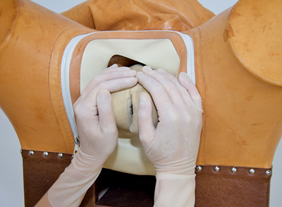 Figure 43a
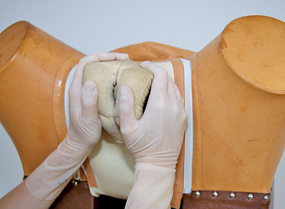 Figure 43b
 Figure 43c
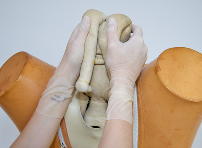 Figure 43d
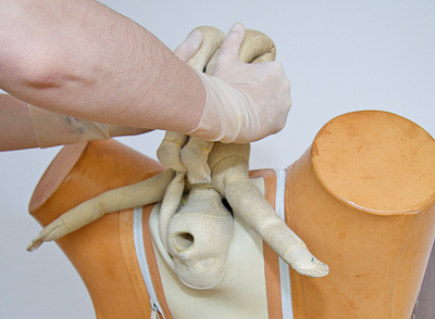 Figure 43e
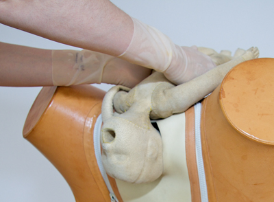 Figure 43f
Procedure
- Never allow a woman to push before there is complete dilation and before the urge to push is present.
- Allow the buttocks to engage as far as possible before active pushing is commenced.
- Make an episiotomy incision at the end of the contraction preceding the contraction in which the birth is expected; this is the case when the buttocks are about to emerge.
- Instruct the woman to push powerfully during the next contraction.
- Make sure that with the birth of the torso the back rotates forwards (ventral side of the mother); if this does not happen spontaneously, the back must be actively rotated to the front.
- Loosen the umbilical cord as soon as the navel has been born to prevent this being placed under tension.
- Wait until the scapular tips are born.
- Apply Bracht’s manoeuvre:
- cover the buttocks (the thumbs splint the femora, the fingers cover the sacrum)
- allow the woman to push powerfully; do not pull, but support the torso until the arms have been born and the hairline is visible
- rotate the child at the height of the posterior hairline around the pubic symphysis towards the mother’s abdomen
- have the woman to push hard during this process
- if necessary apply external expression during this (assistant
- If Bracht’s manoeuvre fails then the birth should be continued with a (partial) breech extraction.
(Partial) breech extraction
It is not always possible to deliver a foetus in the breech presentation with the help of Bracht’s manoeuvre. If the foetus exhibits hypotonia as a consequence of the (analgesic) medications supplied to the mother, or as a consequence of asphyxia, the arms of the foetus will usually be folded around the head during the birth and will not be born spontaneously.
In this case the arms can be delivered using appropriate manoeuvres such as the method described below.
Delivery of the arms
Procedure
- Take the child’s legs in the fork grip with the hand that corresponds to the abdominal side of the child.
- Firmly move the legs ventrally (abdominal side of mother).
- Insert the index and middle finger of the other hand, along the posterior shoulder and upper arm of the child until the elbow fold [Figure 44].
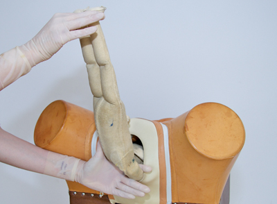 Figure 44
- Move the upper arm of the foetus supported as it were in the gap between the two stretched fingers towards the abdominal side of the child and in so doing sweep it across the child’s face. Do this until the arm has been born.
- Move the child’s feet as far as possible dorsally (back of mother) and repeat the procedure for the other arm.
The anterior arm can also be delivered first; again, the arms should be delivered with the hand that corresponds to the back of the child.
Delivery of the head
For the birth of the head it is important that the occiput is located under the pubic symphysis. Another condition is maximum flexion of the head.
The head can be delivered in various ways, for example with the help of forceps. A manual method is described below.
Procedure
- Take the legs in the fork grip and lift up the torso.
- Place the middle finger of the other hand (the right hand if the small fontanel is located in the right half of the pelvis; the left hand if in the left half) in the mouth of the foetus, the thumb against the lower jaw and the index and ring finger on the upper jaw.
- Have the child rest with the stomach and spread legs on the forearm, in other words with the legs hanging on either side.
- Manoeuvre (rotate) the head if necessary with the inserted fingers, so that the small fontanel comes to lie under the pubic symphysis and at the same time is kept flexed.
- Place two fingers from the other hand around the back of the neck, one on either side, and exert traction on the child’s shoulders [Figure 45].
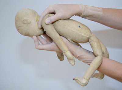 Figure 45
- Important: no traction may be exerted using the finger inserted in the mouth.
- The external hand pulls the child downwards until the posterior hairline is visible.
- Have an assistant exert pressure on the head from above the pubic symphysis, so that less traction needs to be exerted via the neck.
- As the posterior hairline becomes visible, gradually move the torso ventrally so that the head is born.


