Palpation of the aorta takes place in the same way as deep palpation of the abdomen; palpation of the other arteries can best be done using the three middlemost fingertips. Be aware that sometimes one’s own pulse is felt. If in doubt, simultaneously palpate the patient’s carotid artery or auscultate the heart. Take care not to press too hard as, it is very easy to occlude particularly the small arteries in particular!
When palpating the arteries note:
- Size of pulses:
+ present, ± weakly present, – absent
Absent or weak pulses can be indicative of stenosis of the blood vessel or stenosis of a blood vessel closer to the heart which supplies blood to the examined blood vessel. Inability to palpate a dorsal pedal artery does not necessarily signify the presence of pathology. In 10 to 15% of cases the artery is anatomically absent. - Character of the pulse wave
Please refer to the characteristics of the pulse wave as described under the heart examination. - Direction of the pulses
Normally there are transmitted pulses that are only felt on the upstroke and the downstroke. In the case of aneurysm formation (concentric dilatation of a vessel) pulsations can be felt in all directions and are termed expansile pulsations. - Condition of the blood vessel
Normally the peripheral blood vessels are supple and can be easily compressed. In the case of extensive arteriosclerosis, however, the blood vessel can feel tough and hard. - Diameter of the blood vessel
In the case of aneurysm formation the diameter can be markedly increased.
Individual arteries
Common carotid artery [Figure 25]
Palpate both sides at the height of the caudal part of the anterior edge of the sternocleidomastoid muscle. Do not press too hard or palpate both sides at the same time!
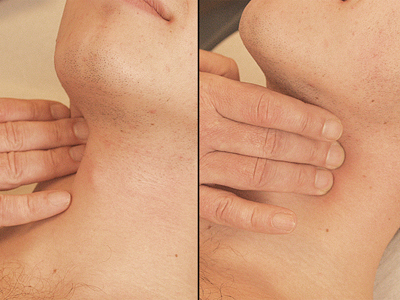 Figure 25: Common carotid artery
Subclavian artery [Figure 26]
Palpate in the supraclavicular space in the caudal direction under the clavicle.
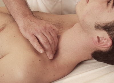 Figure 26: Subclavian artery
Upper extremities
Axillary artery [Figure 27]
Palpate (high) in the armpit, posterior to the attachment of the pectoralis major muscle, towards the humerus. As circulatory disorders of the lower extremities are far more common than those of the upper extremities, the subclavian artery, axillary artery, brachial artery and ulnar artery are not routinely palpated.
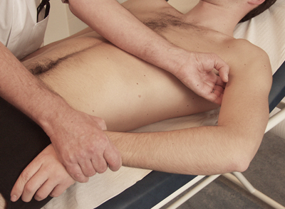 Figure 27: Axillary artery
Brachial artery [Figure 28]
Palpate along the direction of the medial bicipital groove, between the thickest part of the muscle of the biceps and triceps towards the humerus and/or the elbow crease.
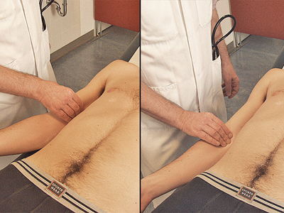 Figure 28: Brachial artery
Radial artery [Figure 29]
Palpate on the ventroradial (thumb) side of the wrist, somewhat proximally to the radial styloid process.
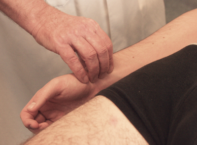 Figure 29: Radial artery
Ulnar artery [Figure 30]
Palpate on the ventral ulnar (little finger) side of the wrist at the height of the ulnar styloid process.
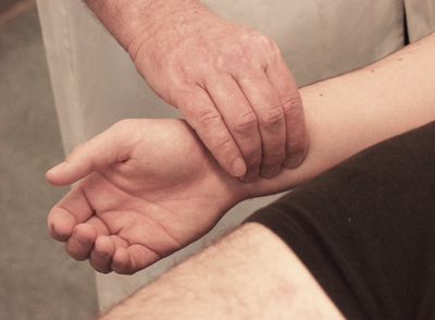 Figure 30: Ulnar artery
Abdomen
Abdominal aorta [Figure 31]
Palpate deep in the epigastrium when the abdominal muscles are relaxed.
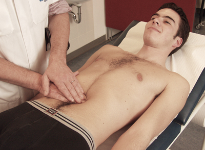 Figure 31: Abdominal aorta
Femoral artery [Figure 32]
Palpate medially in the groin fold with the fingers pointing straight down and parallel to the groin fold. If necessary, once its position has been determined the femoral artery can subsequently be palpated lengthwise.
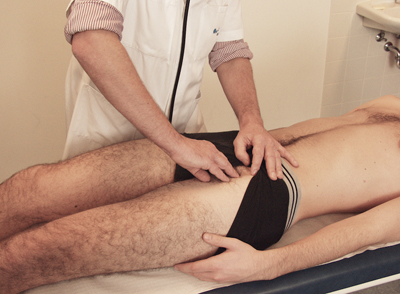 Figure 32: Femoral artery
Lower extremities
Popliteal artery
In supine position: [Figure 33]
Bend patient’s knee at a 45-degree angle and palpate between the fingertips of both hands in the direction of the tibial plateau (in which the thumbs rest on the dorsal edge of the tibial plateau).
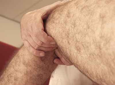 Figure 33: Popliteal artery – In supine position
In prone position: [Figure 34]
Bend patient’s knee at a 45-degree angle; allow it to rest against your shoulder and feel again in the direction of the tibial plateau.
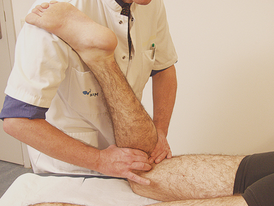 Figure 34: Popliteal artery – In prone position
Posterior tibial artery [Figure 35]
Palpate under and behind the medial malleolus (= tibial malleolus, inner ankle).
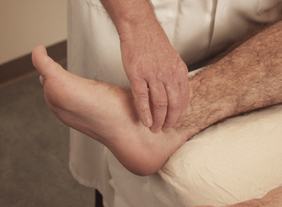 Figure 35: Posterior tibial artery
Dorsal pedal artery [Figure 36]
Palpate the dorsum of the foot just laterally to the tendon of the extensor hallucis longus muscle.
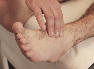 Figure 36: Dorsal pedal artery
Chart – intensity of pulsations [Figure 37]
The intensity of the pulsations can be presented in a chart.
The arteries of the upper extremities have not been included in the overview.
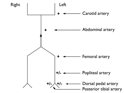 Figure 37: Chart – intensity of pulsations
+ = present
+/- = weakly present
– = absent


