Procedure
- The patient lies in supine position on the examination table.
- During palpation of the dorsal muscles, the patient should be lying on their abdomen.
- Stand next to the patient and palpate with the fingertips.
- Ask the patient to indicate exactly where and when they experience pain.
- In the case of abnormal findings, compare left and right.
- Note:
- Any swelling and its characteristics.
- Muscle tone (fingers transverse to the direction of the fibres).
- Abnormal mobility.
- Abnormal structures.
- Discontinuity.
- The following bone parts are accessible for palpation:
- Greater trochanter (palpate preferably during passive endorotation of a slightly flexed hip) [Figure 33].
- To detect right/left asymmetry (collum fracture, coxa vara, abduction contracture), determine the position of both greater trochanters with respect to both anterior superior iliac spines.
- For the aforementioned abnormalities, this distance will be smaller on the affected side.
- Estimate this distance by placing the thumbs on both anterior superior iliac spines and the index fingers on both greater trochanters (if necessary, mark both these structures with a pencil) [Figure 34].
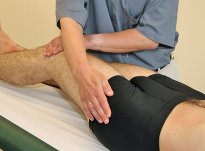 Figure 33
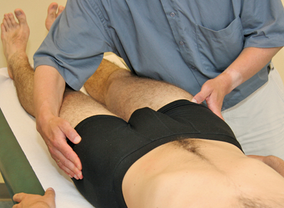 Figure 34
- Symphysis and superior pubic ramus [Figure 35] (symphysiolysis and tendonitis of the pectineus muscle).
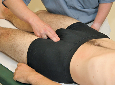 Figure 35
- Ischial tuberosity (palpate at 90° flexion of the hip in prone position) (hamstrings tendonitis) [Figure 36].
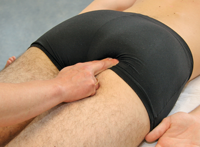 Figure 36
The following soft tissues are accessible for palpation:
- Skin: Palpate mainly with the back of the hand.
- Muscles: Most of the muscles stated in the previous section are accessible for palpation (palpation of the tone of the rectus femoris muscle) [Figure 37].
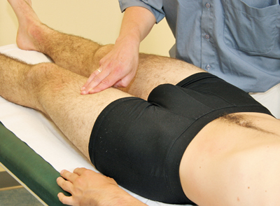 Figure 37
- Bursitis: The trochanteric bursa (between the greater trochanter and the iliotibial band) can only be palpated in the case of swelling. The overlying iliotibial band hinders palpation. Any tenderness should be noted.
- Nerves: Femoral nerve (in the groin) (neuritis); Valleix points (palpate in prone position) [Figures 38, 39] (sciatic-like symptoms).
- Valleix Points: Palpate the points at L5 (paravertebral) and the middle of the maximum curvature of the gluteus maximus muscle and the middle of the gluteal fold.
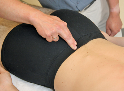 Figure 38
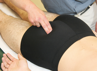 Figure 39
- Blood Vessels: Femoral artery (in the groin): claudication, aneurysms.
- Lymph Glands:
- Horizontal groin glands (in the groin around the inguinal ligament) (Poupart).
- Vertical groin glands (in the groin region perpendicular to, and under, the inguinal ligament).
When indicated, determine whether there is:
- Pain upon axial compression. With the ulnar side of a clenched fist, apply force against the heel in the axial direction with extended hip and knee [Figure 40] (collum fracture).
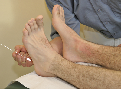 Figure 40
- Tenderness In The Groin: With the palm of the hand, apply considerable compression in the ventro-dorsal direction [Figure 41] (coxarthrosis, collum fracture).
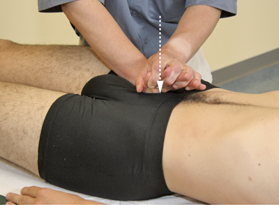 Figure 41
- Trochanter Tenderness: Exert simultaneously from the lateral sides at the height of both greater trochanters considerable compression with the palms of the hands in the direction of the symphysis [Figure 42] (collum fracture, pelvic fracture, symphysiolysis).
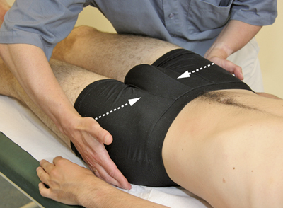 Figure 42


