Anatomy Of The Foot
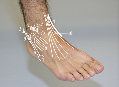 Figure 120a: Lateral Side Of The Right Foot
- Tuberosity of fifth metatarsal.
- Anterior talofibular ligament.
- Calcaneofibular ligament.
- Peroneal trochlea.
- Tendon of the peroneus brevis muscle.
- Tendons of the extensor hallucis longus muscle and extensor digitorum longus muscle.
- Tendon of tibialis anterior muscle.
- Muscle belly of extensor digitorum brevis muscle.
- Tendons of the peroneus longus and peroneus brevis muscles.
- Talonavicular joint.
X = Sinus Tarsi, O = Head of Talus (lateral part)
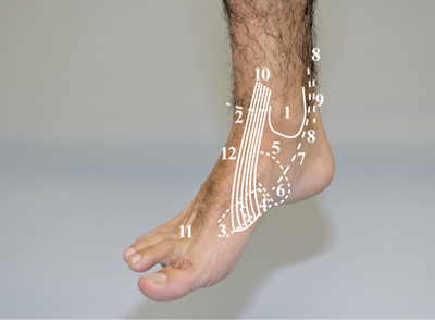 Figure 120b: Medial Side Of The Right Foot
- Medial malleolus.
- Ventral talocrural joint space.
- First metatarsal bone.
- Medial cuneiform bone.
- Head of talus (medial part).
- Tuberosity of navicular bone.
- Sustentaculum tali.
- Tendon of tibialis posterior muscle.
- Tendon of the flexor digitorum longus muscle.
- Tendon of tibialis anterior muscle.
- Tendons of the extensor digitorum longus muscle.
- Talonavicular joint.
Procedure
- The patient sits on the examination table with the lower legs dangling down and no weight bearing on the feet.
- If necessary, sit on a stool in front of the patient.
- Palpate with the tips of your fingers.
- Ask the patient to indicate exactly when and where they feel pain.
- In the case of an abnormal finding compare left and right.
- Note:
- Any swelling and characteristics.
- Muscle tone (fingers perpendicular to the course of the fibres).
- Abnormal mobility.
- Abnormal structures.
- Discontinuity.
- The following bones and joints are accessible for palpation:
- Anterior border of tibia (fracture, subperiosteal haematomas) [Figure 121].
- Medial tibial fascia (shin splints) [Figure 121].
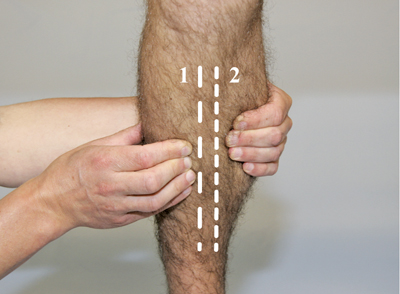 Figure 121
Palpation Of The Tibia:
1. Anterior border (partly indicated).
2. Medial fascia (partly indicated).
In this position the skin is slightly stretched by the palpating fingers to allow the contours of the anterior border to be visualised more clearly.
- Medial malleolus (fracture).
- Distal fibular shaft (fracture).
- Lateral malleolus (fracture).
- Talocrural joint: This joint can be palpated ventrolaterally and behind the medial and lateral malleolus, if necessary during alternating dorsal and plantar flexion (osteophytes) [Figure 122]
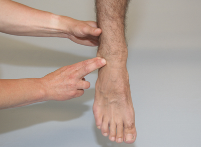 Figure 122
Grip for palpation of the ventrolateral talocrural joint space and the trochlea of the talus (foot is in plantar flexion).
- Subtalar joint and talonavicular joint (subtalar joint can be palpated indirectly at the height of the sinus tarsi, the joint space itself cannot be felt) [Figure 123].
- The talonavicular joint is partially palpable in the inversion position, after the lateral part of the head of the talus has been located. The relevant joint space will be located mediodistally from this [Figure 124].
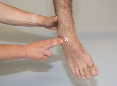 Figure 123
× = Talonavicular Joint
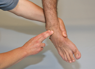 Figure 124
- Trochlea of the talus (in plantar flexion) (exostoses) [Figure 122].
Grip for palpation of the head of the talus (lateral part).
The examiner’s left hand attempts to bring the foot into inversion while the right hand stabilises the lower leg slightly and palpates with the fingers mediodistally from the sinus tarsi.
- Head of the talus (lateral part in inversion [Figure 124], medial part in eversion [Figure 125], exostoses).
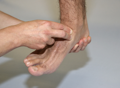 Figure 125
- Calcaneus (Achilles tendon insertion, exostoses, plantar fasciitis).
- Navicular bone (protruding navicular bone in the case of pes planovalgus or an accessory navicular bone) [Figure 126].
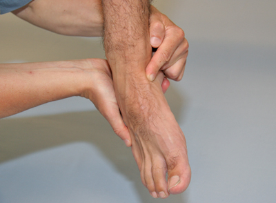 Figure 126
- Cuboid bone (fracture) [Figure 127].
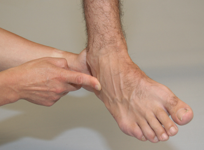 Figure 127
- Cuneiform bone (Lisfranc fracture dislocation) [Figure 128].
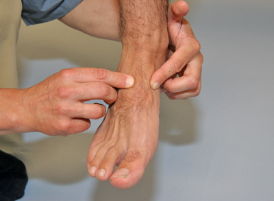 Figure 128
- First to fifth metatarsal bones (fractures).
- Tuberosity of fifth metatarsal bone (avulsion fracture peroneus brevis muscle and possibly peroneus tertius muscle) [Figure 129].
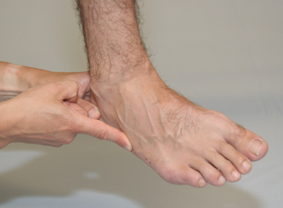 Figure 129
- First to fifth metatarsophalangeal joints (dislocations, rheumatoid arthritis, pes planotransversus, bursae).
- Phalanges (fractures).
- First to fifth proximal and distal interphalangeal joints (hammer toes, clawed toes, curved toes).
- Interphalangeal joint I (dislocation).
If a fracture is suspected, it is important to determine whether pain upon axial compression can be induced. To test this, stabilise the structure to be examined with one hand and with the other hand exert distal pressure along the longitudinal axis of the structure concerned. If the patient now indicates pain where the fracture is suspected, it is referred to as a positive pain upon axial compression [Figure 130].
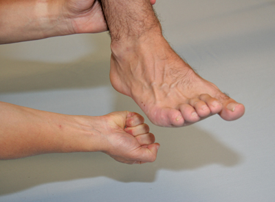 Figure 130
The following soft tissues are accessible for palpation:
- Skin (arteriosclerosis, hyperhidrosis).
- Muscles: Majority of the muscles referred to in the previous section are accessible for palpation (palpation of the tone of the gastrocnemius muscle and palpation of dimples and herniations of the gastrocnemius muscle).
- Tendons:
- Tendon of the extensor digitorum longus muscle, peroneus tertius muscle, extensor hallucis longus muscle and tibialis anterior muscle at the height of the retinaculum of the extensor and distally from this site.
- Tendon of the peroneus longus and peroneus brevis muscles at the trochlea peronealis [Figure 131] and also anterior to the peroneus brevis and tertius muscles at the tuberosity of the fifth metatarsal bone [Figure 129].
 Figure 131
- Tendon of the tibialis posterior muscle, flexor digitorum longus muscle and flexor hallucis longus muscle at the height of the sustentaculum tali [Figure 132] and dorsally and proximally to the medial malleolus, Achilles tendon proximally to the calcaneus (dorsal), plantar fascia at the calcaneal tuberosity site of attachment (plantar) [Figure 133].
If necessary, the tendons may be palpated during movements to determine whether there are crepitations (crepitations of the tendon sheath).
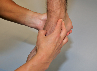 Figure 132
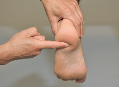 Figure 133
- Ligaments:
- Anterior talofibular ligament (lateral) [Figure 134].
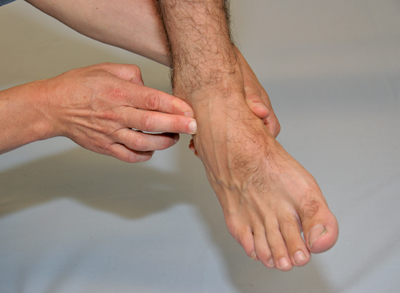 Figure 134
- Calcaneofibular ligament (lateral) [Figure 135].
- Deltoid ligament (medial).
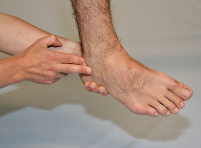 Figure 135
- Nerves:
- Digital nerves (Morton’s metatarsalgia or Morton’s neuroma) – palpate for tenderness [Figure 136].
- ‘Points of Impingement’ in the tarsal tunnel and in the distal continuation of the branches (medial and lateral plantar nerves) of the tibial nerve and at the retinaculum of the extensors, mostly immediately lateral to the extensor hallucis longus muscle (deep peroneal nerve) – check for throbbing and radiating pain.
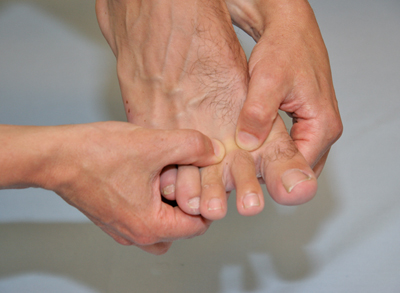 Figure 136
Grip for palpation of digital nerves. Here as an example only the palpation of the digital nerves between the first and second metatarsal bones, and between the second and third metatarsal bones is illustrated.


