Required Instruments
- Head lamp [Figure 32].
- Gloves.
- Tongue spatula.
- Lidocaine spray 10%.
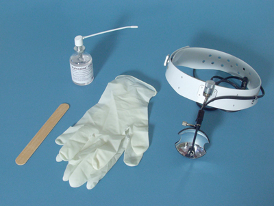 Figure 32
Examination Technique
- For the examination of the mouth and the oropharynx, the patient sits on a chair with adequate back support.
- Sit on a height-adjustable stool beside or in front of the patient.
- Your eyes should be at the same height as the patient’s mouth.
- Wear gloves when palpating the structures inside the oral cavity.
- Before starting the examination run the gloves under a tap to make them wet.
Procedure
- Ask the patient to keep their head straight.
- Ask them to open their mouth wide, keeping the tongue on the floor of the mouth.
- Shine the light on the pharyngeal arches.
- You are now able to inspect the palatal arches and the uvula.
- Pay particular attention to the symmetry of the palatal arches and the position of the uvula. This should be positioned in the middle and hang straight down.
- For this inspection, you can hold the tongue down with a spatula if necessary, by pressing on the front two-thirds of the tongue. With the tongue inside the mouth as described above, you will not hurt the patient, because the tongue is not pressed onto the teeth. You will also not touch the gag reflex site on the back of the tongue [Figure 43].
- Ask the patient to say “ah” and check if the palatal arches move upwards symmetrically and whether the uvula stays in the middle.
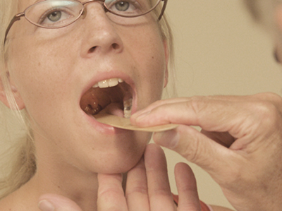 Figure 43
- Aim the light onto the tonsillar fossae, which are located bilaterally, between the palatoglossal arch and the palatopharyngeal arch.
- In children, you will nearly always be able to detect the tonsils. It is important that they are not inflamed, i.e. showing no signs of infection (redness and coating). If they are visible in an adult who does not have a cold, but who does complain of general malaise, a thorough examination of at least the head and neck lymph node stations is indicated.
- Finally, inspect the posterior oropharyngeal wall.
- Assess the colour and presence of hyperaemia.
- In patients with a severe cold, you will be able to see mucus running down from the nose.
- In children with grossly enlarged adenoids, lymphoid tissue can occasionally be visible on the posterior pharyngeal wall as a continuation of the adenoids.
- Again, you should check for any swellings on the posterior pharyngeal wall.
- If necessary, these can be palpated following administration of an anaesthetic (lidocaine spray) to the entrance and the posterior wall of the oropharyngeal area.
- Describe the swelling in more detail.


