The left ventricle has a point that beats against the chest. This phenomenon is termed the apex beat. Due to the heart apex lying close to the thoracic wall in the left semi-prone position, the apex beat is more clearly palpable in this position. Therefore, the position of the apex beat should be determined in the supine position. The other characteristics of the apex beat should be determined in the left semi-prone position.
In a significant number of adults, the apex beat cannot be palpated in the supine position. Thus, it will be impossible to determine the location of the apex beat. In such cases, the location of the apex beat and its characteristics can sometimes be assessed in the semi-prone position.
Locating The Apex Beat
The patient is initially examined in the supine position. Normally, the apex beat is located in the 4th or 5th intercostal space on/within the midclavicular line. If the apex beat is located more to the left, then the heart is enlarged (cardiomegaly) or displaced.
Place a hand on the left half of the thorax with the fingers parallel to the intercostal spaces. The breasts should be carefully pushed upwards when examining female patients. Use the flat of the hand to gain a general impression of the overall location of the apex beat. Use 2 or 3 fingertips to determine the position of the apex beat more accurately. [Figures 5, 6].
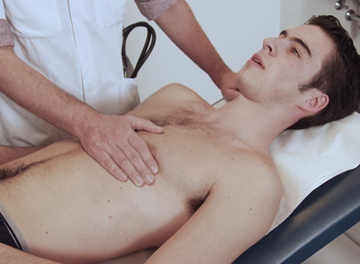 Figure 5: Locating The Apex Beat (1)
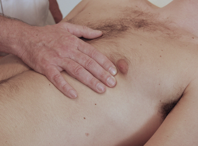 Figure 6: Locating The Apex Beat (2)
Duration Of The Apex Beat
Upon determining the location of the apex beat, examine the other characteristics in the left semi-prone position. [Figure 7]. The impulse of the apex beat should not last any longer than one-third of systole. If the impulse persists for longer, it is referred to as sustained. This is indicative of left ventricular pressure overload (concentric left ventricular hypertrophy). Palpate using the flat of the hand and then the fingertips. The duration of the apex beat compared to the duration of systole is estimated by simultaneously palpating and auscultating (systole is located between the 1st and 2nd tone).
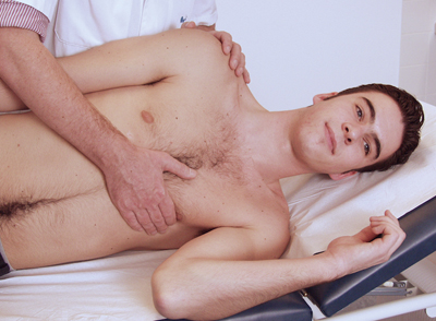 Figure 7: Assessing The Characteristics Of The Apex Beat
Area Of The Apex Beat
Assess the size of the apex beat in the left semi-prone position using the flat of the hand and follow with fingertips. Normally, the diameter of the apex beat area lies within just one intercostal space. If the apex beat can be palpated over more than one intercostal space, it is known as a displaced apex beat. A displaced apex beat is indicative of ventricular dilatation or cardiac aneurysm formation.
Amplitude Of The Apex Beat
In the left semi-prone position, use the fingertips to estimate the intensity of the apex beat as normal, strong or weak. A strong apex beat can be associated with pressure overload (aortic stenosis), volume overload (mitral insufficiency) or hyperdynamic circulation (anaemia). A weak apex beat can be found in the event of hypovolaemia (shock as a consequence of underfilling of the vascular system), pericardial effusion (fluid in the heart sac) or constrictive pericarditis (inflammation of the heart sac as a result of which cardiac contraction and expansion is impaired).
Composition Of The Apex Beat
Repeat palpation using the fingertips, with the patient in the left semi-prone position. Assess whether the apex beat consists of a single impulse or whether there is an extra impulse before the strong impulse. The latter is referred to as a double (bifid) apex beat and is always pathological. It is caused by increased atrial emptying, which cannot normally be felt, but in this case, can be palpated. This occurs with left atrial volume or pressure overload (mitral insufficiency or left ventricular hypertrophy).


