Required Instruments
- Head light [Figure 21].
- Hartmann nasal speculum.
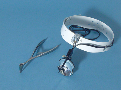 Figure 21
Examination Technique
- For all examinations of the nose, the patient sits on a chair which provides adequate back support.
- Sit on a height-adjustable stool beside or in front of the patient.
- Your eyes should be at the same height as the patient’s nose.
Procedure
- Ask the patient to hold their head slightly backwards so that the nostrils and the columella become visible.
- Aim the light from the head lamp in the nostril that is to be examined.
- Hold the nasal speculum with your hand in a neutral position, between pronation and supination.
- One leg of the speculum should lie in the palm of your hand in the groove between the thenar and hypothenar eminences [Figure 28].
- Place your middle, ring, and little fingers against the other leg of the speculum.
- The index finger should lie against the speculum blade [Figure 29].
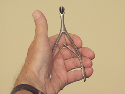 Figure 28
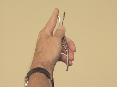 Figure 29
- Place the four fingers of your other hand on the patient’s forehead and press the nasal tip slightly upwards using the thumb [Figure 30].
- This allows you to properly assess the position of the columella and the frontal part of the nasal septum.
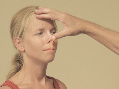 Figure 30
- Insert the closed nasal speculum with the blade parallel to the nasal floor or with the opening at a 45° angle relative to the nasal floor [Figure 31].
- If necessary, use the thumb of your other hand to guide the speculum, to avoid touching the blood vessels of the Kiesselbach plexus or the nasal septum.
- If the tip of the nasal speculum has passed the nasal valve area, carefully open the speculum.
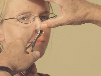 Figure 31
- The inferior, and occasionally the middle, turbinate are now visible.
- Describe the mucosa covering these structures (smoothness, colour, swelling), plus the presence of any pus, mucus or blood.
- The inferior and middle meatus should also be visible now and can be assessed for the presence of pus, mucus, blood or a foreign object.
- If the nasal mucosa are swollen to such an extent that the middle turbinate and meatus cannot be inspected, repeat the examination once you have decongested the nasal mucosa.
- Once you have finished the examination, remove the nasal speculum in a slightly open position from the patient’s nose.
- In this manner, you avoid trapping and removing any nasal hairs, which can be painful.
- Clean the speculum with soap and water.
In very young children, anterior rhinoscopy can be carried out more easily using an otoscope with a 6mm ear speculum attached.


