Required Instrument
- Head light.
Examination Technique
- For all examinations of the nose, the patient sits on a chair which provides adequate back support.
- Sit on a height-adjustable stool beside or in front of the patient.
- Your eyes should be at the same height as the patient’s nose.
Procedure
- Ask the patient to hold their head up straight and aim the light of the examination lamp or head light onto the patient’s nose.
- Describe the colour and smoothness of the covering skin.
- Assess the position of the nose and look for any visible swellings.
- Ask the patient to hold the head slightly backwards so that the nostrils and the columella become visible.
- Check whether the nasal columella is straight.
- Press the tip of the nose slightly upwards with your thumb [Figure 22].
- The frontal part of the nasal vestibulum will now become visible.
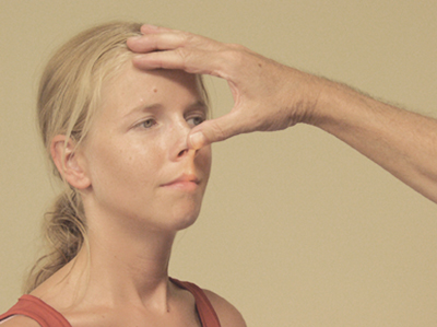 Figure 22
- Check if there is any pus, mucus, blood or possibly a foreign object visible in the nose.
- In this position, occasionally the inferior turbinate and nasal cavity will be visible.
- Assess the colour and any swelling of the turbinate mucosa and accessibility of the nasal cavity.
- Again, check for the presence of pus, mucus, blood, or a foreign object.
- Take hold of the nasal bone between thumb and index finger [Figure 23] and move the covering skin carefully over the bony part of the nasal skeleton.
- Note any swellings and other irregularities on the surface that may indicate recent or old damage to the nasal bone.
- Ask the patient whether this is painful.
- Whilst feeling the nose with your fingers in this manner, palpate towards the nasal tip.
- For any swellings that you may come across, describe the size, perimeters, surface, consistency, and mobility in relation to the surrounding structures.
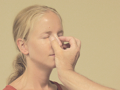 Figure 23
- Finally, assess the accessibility of the nose.
- To do this, first close off one nostril and then the other by placing a finger on the underside of the nostril [Figure 24].
- Make sure to wear a glove.
- Then ask the patient to inhale and exhale through the opened nostril.
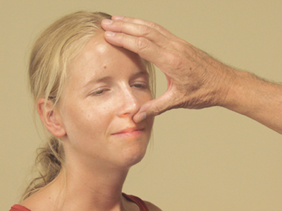 Figure 24


