These tests may be indicated in a patient with lower back symptoms and an abnormal posture without rigid or structural components.
Procedure
Test in a supine or prone position, the clinically most relevant pelvic muscles for strength.
These are:
- Erector muscle of spine (see “muscle tests”).
- Rectus abdominis muscle (see “muscle tests”).
- Hip flexors (iliopsoas muscle and rectus femoris muscle in particular) in supine position. Allow the patient to lift the leg, flexed at hip and knee, to the torso and provide opposite force at the level of the ventral side of the upper leg [Figure 60].
- Hip extensors (the hamstrings or ischiocrural muscles in particular) in supine position. Allow the patient to move the heel of the bent leg to the buttock and provide an opposite force at the level of the dorsal side of the lower leg [Figure 61].
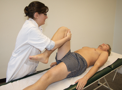 Figure 60
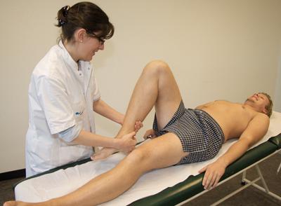 Figure 61
Interpretation
For the erector muscle of the spine and the rectus abdominis muscle, see “muscle tests.” For the hip extensors and the hip flexors, the strength can be graded on a scale of 0 to 5. Grade 5 is applied in a person with normal strength.
With the patient in supine or prone position, test the length of the following muscles:
- Erector muscle of spine. Bend forward whilst seated with legs stretched, touching the toes [Figure 62].
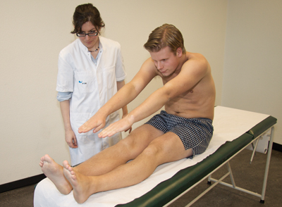 Figure 62
Interpretation
During these tests, the length of the hamstrings and the triceps surae are also assessed. Therefore, various variations may be possible when the patient bends forward. With a normal length of the erector muscle of spine, there will be an increase in the convexity of the spine to the front and in the lumbar region a flattening of the lordosis. With normal length hamstrings, the pelvis will tilt forward and the legs will not bend in the knees. With normal length triceps surae muscle, the legs will not bend in the knees and the toes will continue to point upwards (= midpoint between dorsal and plantar flexion).
- Hip flexors. In supine position with lower legs hanging down, allow the patient to alternately bring the bent legs to the chest (both hands placed in the back of the left or right knee, respectively) [Figure 63].
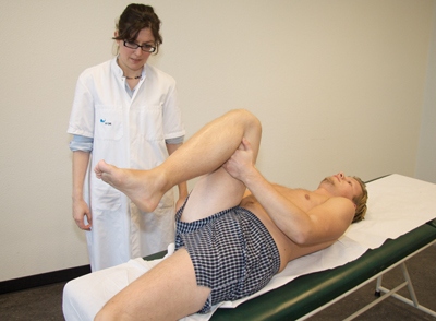 Figure 63
Interpretation
When the bent leg is pulled to the chest, the contralateral leg should remain in the same hanging position. Should the upper contralateral leg come off the examination table, it indicates an iliopsoas muscle which is too short. If the contralateral leg stretches in the knee, it indicates a too short rectus femoris muscle. Both phenomena can also occur simultaneously.
- Hip extensors (performance of this test is determined by the presence or absence of shortness of the hip flexors).
A. If hip flexors are not shortened.
The examiner lifts the stretched leg of the patient, whilst the contralateral leg is fixed in a stretched position on the surface. The lumbar lordosis is automatically flattened due to the backwards tilting of the pelvis [Figure 64].
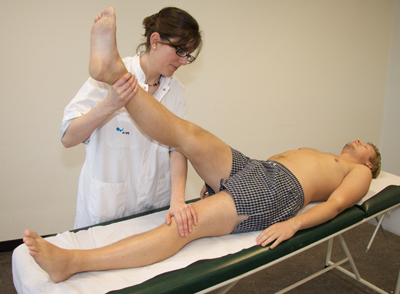 Figure 64
Interpretation
With normal length hamstrings, the lifted leg will form a minimum angle of 80° with the surface.
B. If hip flexors are shortened.
The examiner lifts the stretched leg of the patient, whilst the contralateral leg is bent in the knee and the hip by means of a pillow underneath the knee. The shortening of the hip flexors can then be compensated and the lumbar lordosis is given the opportunity to flatten [Figure 65].
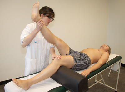 Figure 65
Interpretation
Hip flexors that are too short, prevent the backward tilting of the pelvis during fixation of the leg with the extended hip. Therefore, the test mentioned in A cannot be conducted reliably. The ischiocrural muscles can namely appear shorter than they actually are. With normal length hamstrings the lifted leg will form an angle of 80° with the surface.
C. See test for length of the erector muscle of spine (bend forward whilst seated with legs stretched and trying to touch the toes with the fingers).
- Rectus abdominis muscle. This muscle cannot be optimally tested for length.
General Interpretation
- The stronger the muscle, the bigger the chance of shortening the muscle in question.
- The weaker the muscle, the bigger the chance of lengthening the muscle in question.
- The starting point is that the pelvis, due its potential to tilt, can be seen as a balance.
With the pelvis portrayed as a balance, the erector muscle of spine and the hip flexors and the rectus abdominis muscle and hip extensors are agonists of each other.
With a muscular imbalance at this level, muscles are shortened or lengthened. For example, exaggerated lumbar lordosis may be the result of a too strong erector muscle of spine and a weak rectus abdominis muscle. Management should be aimed at lengthening the overly strong muscles and strengthening the overly long muscles, both with aid of exercise therapy (stretch exercises and muscle strengthening exercises, respectively).


