Murmurs over the large central and peripheral arteries can be indicative of constrictions in either the blood vessel or the supplying blood vessel located closer to the heart.
Note the following during auscultation of the arteries:
- continually compare left and right
- listen with the diaphragm of the stethoscope; murmurs are high-frequency
- do not press too hard; this can lead to a murmur due to artificial constriction of the vessel
- record the findings in the chart with the notation S+ of S-. [Figure 37]
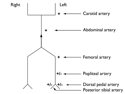 Figure 37
Specific auscultation locations
Common carotid artery [Figure 38]
To prevent confusion have the patient briefly hold their breath.
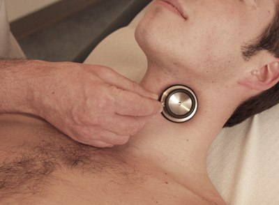 Figure 38: Common carotid artery
Subclavian artery [Figure 39]
In the case of small, slender patients it can be difficult to obtain sufficient skin contact with the stethoscope.
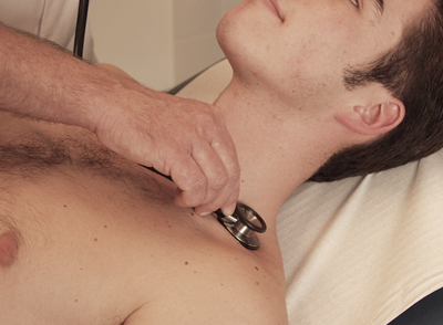 Figure 39: Subclavian artery
Abdominal aorta [Figure 40]
Listen both above the navel and at the height of the navel (at the bifurcation).
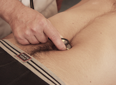 Figure 40: Abdominal aorta
Renal artery [Figure 41]
Listen on both sides of the aorta halfway between the lowest point of the sternum and the navel.
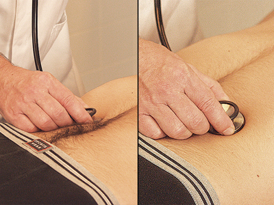 Figure 41: Renal artery
External iliac artery [Figure 42]
Listen to the section between the navel and the femoral artery.
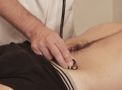 Figure 42: External iliac artery
Femoral artery [Figure 43]
Listen on both sides medially in the groin fold at the point where the femoral artery was felt by means of palpation.
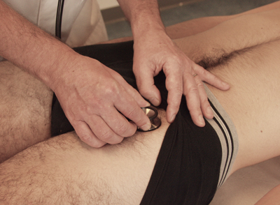 Figure 43: Femoral artery


